Retinal detachment is a serious eye condition that requires immediate medical attention. In this article, we will explore the latest advances in retinal detachment treatments, including both traditional methods and recent developments. We will also discuss the future of retinal detachment treatments and potential breakthroughs in the field.
Understanding Retinal Detachment
Before diving into the latest treatments, it is important to understand what retinal detachment is and how it occurs. The retina is a layer of tissue located at the back of the eye that is responsible for converting light into signals that the brain can interpret as visual images. Retinal detachment occurs when the retina separates from its underlying supportive tissue.
Looking ahead, the future of retinal detachment treatments holds great promise, with potential breakthroughs in retinal repair, the role of technology, and the development of non-invasive treatments.
This condition can lead to vision loss if not treated promptly. It is essential to recognize the causes and symptoms early on to seek appropriate medical care.
What is Retinal Detachment?
Retinal detachment happens when the retina separates from the supportive tissue beneath it. This separation disrupts the blood supply to the retina, leading to decreased oxygen and nutrient delivery. Without proper blood flow, the retinal cells can become permanently damaged.
Imagine the retina as a delicate piece of fabric, intricately woven with specialized cells that capture and transmit visual information. When retinal detachment occurs, it’s like a tear in this delicate fabric, causing a disruption in the visual process. The retina, no longer firmly attached to its supportive tissue, becomes vulnerable and starved of the vital resources it needs to function optimally.

Causes and Symptoms of Retinal Detachment
Retinal detachment can occur due to various factors. Some of the common causes include age-related changes, trauma to the eye, severe nearsightedness, and certain eye diseases like diabetic retinopathy. One of the most common symptoms of retinal detachment is the sudden onset of floaters in the field of vision.
Floaters are tiny specks or cobweb-like shapes that seem to drift across your field of vision. They can be quite alarming, especially when they appear out of nowhere. These floaters are actually small bits of debris floating in the jelly-like substance called the vitreous humor, which fills the inside of the eye. When the retina detaches, it can cause these floaters to become more noticeable, as if they are dancing in front of your eyes.
Other symptoms may include flashes of light, a shadow or curtain appearing in the peripheral vision, or a decrease in vision. These visual disturbances can be frightening and should not be ignored. It is crucial to bring any of these signs to the attention of an eye specialist immediately, as early intervention can greatly improve the chances of successful treatment.
Traditional Methods of Treating Retinal Detachment
Retinal detachment is a serious condition that requires prompt treatment to prevent permanent vision loss. Over the years, several traditional methods have been developed to treat this condition, each with its own unique approach and benefits. These methods aim to reattach the retina and restore its normal function, giving patients a chance to regain their vision and quality of life.
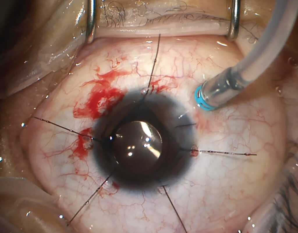
One of the most commonly used techniques for treating retinal detachment is scleral buckling surgery. This procedure involves placing a silicone band or sponge around the eye to indent the wall of the eye and reposition the detached retina. By creating this indentation, the retina is brought into contact with its supporting tissue, facilitating reattachment. Scleral buckling surgery is usually performed under local anesthesia, ensuring patient comfort throughout the procedure. Read more about anesthesia on https://www.nigms.nih.gov/education/fact-sheets/Pages/anesthesia.aspx
The success rate of scleral buckling surgery varies depending on the severity and location of the detachment, but it has proven to be an effective method in many cases. This technique has been refined over the years, with advancements in surgical instruments and techniques, leading to improved outcomes and patient satisfaction.
Scleral Buckling Surgery: A Time-Tested Approach
Since its introduction, scleral buckling surgery has stood the test of time as a reliable and effective method for treating retinal detachment. The procedure has been performed on countless patients, with remarkable success rates in reattaching the retina and restoring visual function. Surgeons who specialize in retinal detachment have honed their skills in performing this intricate procedure, ensuring optimal outcomes for their patients.
Another traditional method for treating retinal detachment is pneumatic retinopexy. This less invasive procedure is typically performed in the office setting, offering convenience and comfort to patients. During pneumatic retinopexy, a gas bubble is injected into the vitreous cavity of the eye. The patient’s head is then positioned in a specific way to allow the bubble to press against the detached retina, effectively sealing the tear or hole.
Pneumatic retinopexy has shown positive outcomes, particularly for certain types of retinal detachments. However, it may not be suitable for all cases and requires careful patient selection and monitoring. The success of this procedure relies on the patient’s ability to maintain the correct head position, allowing the gas bubble to exert its therapeutic effect on the retina. With proper patient education and follow-up care, pneumatic retinopexy can be a valuable treatment option for retinal detachment.
Vitrectomy: A Surgical Breakthrough
Vitrectomy is another traditional method used to treat retinal detachment. This surgical technique involves removing the vitreous gel from the eye and replacing it with a saline solution. By doing so, the surgeon gains direct access to the retina, allowing for any necessary repairs to be made, such as removing scar tissue or sealing retinal tears.
Vitrectomy has been a significant advancement in the treatment of retinal detachment. It offers the advantage of allowing better visualization and manipulation of the retina, leading to improved surgical outcomes. With the aid of specialized instruments and advanced imaging technology, surgeons can perform delicate maneuvers with precision, ensuring the best possible results for their patients. Click here to find more about precision.
As with any surgical procedure, vitrectomy carries some risks and potential complications. However, advancements in surgical techniques, anesthesia, and post-operative care have significantly reduced these risks, making vitrectomy a safe and effective treatment option for retinal detachment.
In conclusion, traditional methods of treating retinal detachment, such as scleral buckling surgery, pneumatic retinopexy, and vitrectomy, have proven to be valuable tools in the hands of skilled retinal surgeons. These techniques have revolutionized the field of ophthalmology, allowing patients with retinal detachment to regain their vision and improve their quality of life. With ongoing research and advancements in technology, the future holds even more promising treatments for this sight-threatening condition.
Recent Developments in Retinal Detachment Treatments
Advancements in medical technology have brought forth significant developments in the treatment of retinal detachment. These innovations aim to enhance surgical techniques, refine laser therapy, and improve medication options for patients.
Innovations in Surgical Techniques
Surgical techniques for retinal detachment have evolved significantly over the years. Minimally invasive procedures, such as robotic-assisted surgery, have emerged as a promising option. Robotic systems can provide surgeons with enhanced dexterity and precision during delicate surgeries, resulting in improved patient outcomes.
Moreover, the integration of artificial intelligence (AI) algorithms into surgical robots has further revolutionized the field. These algorithms can analyze real-time data from the patient’s eye, assisting surgeons in making informed decisions during the procedure. By combining the expertise of surgeons with the analytical capabilities of AI, the success rates of retinal detachment surgeries have soared to new heights.
Additionally, the development of customized surgical tools and imaging technologies has allowed surgeons to optimize their approach based on individual patient characteristics, leading to better surgical success rates and faster recovery times. These tools and technologies enable surgeons to precisely visualize the affected area and plan their surgical strategy accordingly.

Advancements in Laser Therapy
Laser therapy has been a valuable tool in the treatment of certain retinal disorders, including retinal detachment. Recent advancements in laser technology have allowed for more targeted and precise treatment, minimizing damage to surrounding tissue.
Furthermore, the integration of imaging systems with lasers has improved the accuracy of laser therapy. These systems provide real-time feedback, guiding the surgeon’s movements and optimizing treatment delivery. The combination of laser therapy and imaging technologies has the potential to revolutionize the treatment of retinal detachment.
Moreover, researchers are exploring the use of nanotechnology in laser therapy for retinal detachment. Nanoparticles can be engineered to deliver therapeutic agents directly to the affected area, enhancing the effectiveness of the treatment. This targeted approach minimizes side effects and maximizes the therapeutic benefits for patients.
Progress in Medication for Retinal Detachment
While surgery remains the primary treatment for retinal detachment, there have been advancements in medication options that can augment surgical outcomes. Researchers are exploring the use of pharmacological agents that promote retinal healing and prevent further complications.
These medications aim to enhance the reattachment process by stimulating cellular growth and reducing inflammation. Although still in the experimental stages, these pharmacological advancements offer hope for improved treatment outcomes and minimized long-term complications.
Furthermore, gene therapy is emerging as a potential treatment option for retinal detachment. By introducing specific genes into the affected cells, researchers can promote the regeneration of damaged retinal tissue. This innovative approach holds great promise for patients with severe retinal detachment, offering a potential cure rather than just a treatment.
In conclusion, recent developments in retinal detachment treatments have brought about significant advancements in surgical techniques, laser therapy, and medication options. These innovations have the potential to improve patient outcomes, minimize complications, and revolutionize the field of ophthalmology.
The Future of Retinal Detachment Treatments
The future of retinal detachment treatments holds great promise, with ongoing research and development paving the way for potential breakthroughs. Let’s explore some of the areas that researchers and specialists are focusing on.
Potential Breakthroughs in Retinal Repair
Scientists are actively investigating new methods of repairing the retina, including tissue engineering and cell-based therapies. These approaches aim to replace damaged retinal cells with healthy ones, restoring vision in individuals with retinal detachment.
Through the use of stem cells or other regenerative techniques, researchers hope to develop treatments that can regenerate the damaged retinal tissue, potentially revolutionizing the field of retinal detachment treatment in the future.
The Role of Technology in Future Treatments
Technological advances continue to play a vital role in shaping the future of retinal detachment treatments. The integration of artificial intelligence (AI) and machine learning algorithms with diagnostic tools can help physicians accurately detect and classify retinal detachment at an early stage.
Furthermore, the development of novel imaging techniques, such as optical coherence tomography (OCT) and adaptive optics, enables more precise evaluation of retinal structure and function. These technologies aid in treatment planning and monitoring postoperative outcomes, leading to improved patient care.
Predictions for Non-Invasive Treatments
Non-invasive treatments for retinal detachment are a topic of great interest and research. Scientists are exploring alternatives to surgery, such as the use of focused ultrasound or drug delivery systems that target specific regions within the eye.
By developing non-invasive interventions that can achieve retinal reattachment without the need for invasive procedures, researchers hope to minimize patient discomfort, reduce the risk of complications, and improve overall treatment outcomes.
In conclusion, the latest advances in retinal detachment treatments offer hope for improved outcomes and quality of life for individuals affected by this condition.
Traditional methods such as scleral buckling surgery, pneumatic retinopexy, and vitrectomy continue to be effective treatment options.
Recent developments in surgical techniques, laser therapy, and medication options provide further advancements in the field.
It is essential for individuals experiencing symptoms of retinal detachment to seek immediate medical attention and consult with an ophthalmologist to explore the most appropriate treatment options available.

